Title: NEUROSCIENCE - Exploring the Brain
Author: Mark F. Bear, Barry W. Connors, Michael A. Paradiso
Duration: 07/2022-present
Chapter 1 Neuroscience: Past, Present, and Future
- Localization of Specific Functions to Different Parts of the Brain
- Experimental ablation method 实验消融法
- Phrenology 颅相学
- Level of analysis
- Molecular Neuroscience
- Cellular Neuroscience: give neuron their specific functions
- System Neuroscience: neural circuits
- Behavioral Neuroscience
Cognitive Neuroscience: how neural mechanisms responsible for the higher level of human mental activity
- The Scientific Process
- Observation
- Replication
- Interpretation
- Verification
- Further Reading
- All man JM 1999. Evolving Brains
- Clarke E, The human brain and spinal cord
- Corsi P,The Enchanted Loom
- Crick F. 1994, The astonishing Hypothesis: The Scientific Search for the Soul
- Finger S, 1994, Origins of Neuroscience
- Glickstein M. Neuroscience: A Historical Introduction
Chapter 2 Neurons and Glia
Introduction
- Obstacles
- Small size
- uniform, cream-colored appearance under microscope
The Neuron Doctrine
Nissl Strain, Nissle bodies 尼氏体
Nissle bodies are discrete granular structures(颗粒结构) in neurons that consist of rough endoplasmic reticulum(内质网), a collection of parallel, membrane-bound cisternae(膜结合池) studded with ribosomes(散布着核糖体) on the cystosolic surface(细胞质表面) of the membranes(膜).
"Nissl stains"(尼氏染色) refers to various basic dyes(染料) that selectively label negatively charged molecules such as DNA and RNA.
Advantage: 1. distinguishes between neurons and glia; 2. enable histologists to study the arrangement or cytoarchitecture('cyto' -- Greek 'cell') of neurons in different parts of the brain.
- Could not tell the whole stody.
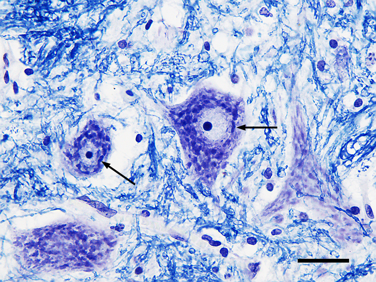
Fig.2.1 - Photomicrograph of Nissl bodies[wiki]
The Golgi Stain 高尔基染色
show neurons have at least two parts: a central region that contains the cell nucleus and numerous thin tubes that radiate away from the central region
- The swollen region(肿) contains
- cell body
- soma(plural: somata) 躯体
- perikaryon(plural: perikarya) 周核体
- The thin tubes
- neurites 神经突: Axibs 轴突 and dendrites 树突
The cell body usually gives rise to a single axon(轴突). The axon is of uniform diameter throughout its length and any branches from it generally extend at right angles -- Axons can extend over great distances in the body.
The Prototypical Neuron
The Soma 躯体
- Spherical central part
Cytosol 胞质溶胶: watery fluid inside the cell; salty, potassium-rich solution; separated from outside by neuronal membrane
- organelles 细胞器: membrane-enclosed structures
The cell body of the neuron contains the same organelles found in all animal cells. Everything contained within the confines of the cell membrane, including the organelles but excluding the nucleus, is referred to collectively as the cytoplasm 细胞质
- Nucleus 细胞核
- Latin 'nut',spherical, central located, 5-10 μm
- nuclear envelope 核膜: Double membrane 0.1μm
- Chromosomes, DNA, Gene
- Gene expression:
- Reading, final product is the synthesis of molecules(Proteins)
- occur in aytoplasm
It is performed by messenger ribonucleic acide/mRNA, the process called transcription 转录(the part of DNA called transcript)(RNA polymerase RNA聚合酶) (Promoter, exon, intron, terminator, transcription factor, RNA slicing RNA剪接)
- translation (protein building blocks: amino acide 氨基酸)
- Neuronal Genes, Genetic Variation and Genetic Engineering
Neurons differ from other cells in the body because of the specific genes they express as proteins
mishaps不幸: gene copy number variations(occur when paternal and maternal DNA mix to create the genome基因组 of the offspring后代); mutation(single nucleotide polymorphisms单核苷酸多态性) knockout mice基因敲除小鼠 knock-in mice 敲入小鼠 transgenic mice 转基因
- Rough Endoplasmic Reticulum 粗面内质网
protein synthesis site in Neurons, free ribosomes(核糖体), polyribosomes 多核糖体(attach by a thread, a single strand of mRNA)
ifference between proteins sythesized on the rough ER and on the free ribosomes: depend on the intended fate of the protein molecule
- inseted into the membrance of the cell or an organelle : Rough ER
- cytosol: free ribosomes
- Smooth Endoplasmic Reticulum 滑面内质网 and The Golbi Apparatus 高尔基体
- without ribosomes
- heterogeneous and perform different function in different locations
- continuous with Rough ER
Golbi apparatus: sorting of certain proteins that are destined for delivery to different parts of the neuron
- The mitochondrion 线粒体
- cristae 嵴
- matrix 嵴内间隙
- cell respiration 细胞呼吸
- Krebs cycle(electron-transport chain) : ADP, ATP cell's energy source
- Nucleus 细胞核
The neuronal Membrane
- barrier
- 5nm, stubbed with protein
- associated with pump substance
- Membrane difference mainly in proteins compositions
The cytoskeleton 细胞骨架
- it gives neuron its characteristic shape
- microtubes
- a straight, thick-wall hollow pipe
- the wall of the pipe consists of the protein tubulin 微管蛋白
- Polymerization 聚合: joining small proteins to form a long strand
microtubule-associated proteins MAPs: participate in the regulation of microtubule assembly(connect wit other parts of the neuron)
Pathological changes 病理变化 in an axonal MAP called tau has been implicated in the dementia失智 that accompanies Alzheimer's decease
microfilaments 微丝: braids of two thin strands that are polymers of the protein actin肌动蛋白
- Neurofilaments 神经丝
- as intermediate dilaments
- most closely resemble bones and ligaments of the skeleton
The Axon
- highly specialized for the transfer of information
- Axon hillock 轴突丘: begin region
No rough ER extends into the axon, and there are few, if any,free ribosomes in the mature axons
- The protein composition is different from soma membrance
All protein in the axonal membrance originate from soma -- enable axonal membrance serve as a wire that sends information over great distance
- Axon collaterals轴突侧支; recurrent collaterals
- axon diameter varies: nerve impluses depends on the axonal diameter.
- The axon terminal: contact with other neurons and pass information(synaps突触)
- Terminal arbor 终端: axons short branches end
- Difference between axon and axon terminal
- microtubules do not extend into the terminal
- the terminal contains numerous small bubbles of membrane(synaptic vesicles突触小泡)
- inside membrance has dense covering protein
- numerous mitochondria -- high energy demand
- The synapse
- presynaptic 突触前: includes axon terminal
- postsynaptic 突触后
- synaptic cleft 突触间隙:space between pre- and post-
- synaptic transformation: pre(electrical) -- cleft(chemical) -- post(electrical)[memory & learing]
- Axoplasmic transport
- wallerian degeneration: the degeneration of axons that occurs when they are cut
fast & slow : kinesin驱动蛋白 'walk' -- fueled by ATP -- move from soma to the terminal [all transport in this direction called anterograde transport 顺行运输]
- [retrograde transport 逆行运输] terminal to soma -- dynein 动力蛋白]
fast anterograde transport was hown by injecting the soma with radioactive amino acides + HRP horseadish pre-oxidase 辣根过氧化物酶
Dendrites 树突
- dendritic tree: the dendrites of a single neuron
- dendritithic branch
- different shapes and sizes are used to classify different groups of neurons
- dendritic spines 树突棘: neurons with specialized structure
Classifying Neurons
- Neural structure
- number of neurites神经突数: unipolar, bipolar, multipolar
- dendrites: stellate cells and pyramidal cells
- whether have spines: spiny and aspinous
- connections: primary sensory neurons, motor neurons, interneurons
- Axon length: Golgi type Ⅰneurons(projection neurons); Golgi type Ⅱ neurons(local circuit neurons)
- Gene expression
Glia 神经胶质
Astrocytes 星形胶质细胞
- most numerous glia, filled most of the space between neurons
- regulate the chemical content of this extracellular space
- special proteins in their membrance actively removes many neurotransmitters from the synaptic cleft
Myelinating Glia 有髓胶质细胞
- oligodendroglial 少突胶质细胞 Schwann cells -- provide layers of membrane that insulate axons
- myelin 髓磷脂
Ohter Non-Neuronal Cells
- ependymal cells 室管壁细胞: cell migration
- microglia 小胶质细胞
- vasculature 脉管系统: arteries, veins, capillaries
Chapter 3 The Neuronal Membrane at Rest
Introduction
- action potential
information is encoded in the frequency of action potentials of individuals neurons as well as in the distribution and number of neurons potential in a given nerve
- excitable membrane
- esting membrane potential/ resting potential
The Cast of Chemicals
Cytosol and extracellular fluid 细胞质和细胞外液
- Water
- he oxygen atom has a great affinity for electrons than does the hydrogen atoms
- oxygen acquires a net negative charge, hydrogen -- positive 这是本身电性
- polar covaleng bonds 极性共价键
- Ions
- Na+: oxygen, Cl-: hydrogen
- spheres of hydration: insulating the ions
- monovalent 单价; divalent 二价; cations 阳离子; anions 阴离子
- Na+,K+; Ca2+; Cl- s
The Phospholipid Membrane 磷脂膜
- phospholipid has long nonpolarchains of carbon atoms bonded to hydrogen atoms
- pholipid has a polar phosphate group attched to one end of the molecule
- one head is hydrophilic 亲水的(head), the other is hydrophobic 疏水的
- Two molecules thick, phospholipid bilayer 磷脂双分子层
Protein
- enzymes: catalyze chemical reactions in the neurons
- cytoskeleton: special shape
- receptors 受体: sensitive to neurotransmitters神经递质 are all made up of protein molecules
amino acid氨基酸(central carbon group/the alpha carbon: an amino group+a carboxyl group+R group)*20 kinds -- peptide bonds 肽键
Primary structure -- alpha helix 阿尔法螺旋 -- tertiary strucutre 三级结构(bend fold complex 2D structure) quaternary structure 亚基
- Channel Protein:Ion channels, ion selectivity, gating, ion pumps
The Movement of Ions
- Diffusion: concentration gradient+membrane has channels permeable to the ions.
- Electricity: conductance: if conductance is zero, there will be no current.
The Ionic Basis of the Resting Membrane Potential
- $V_m$: Membrane potential(neuronal membrane at any moment).
- microelectrode: thin glass tube with an extremely fine tip.
Equilibrium potentials
Ionic equilibrium potential: the electrical potential difference that exactly balance an ionic concentration gradient.
- Large changes in membrane potential are caused by minuscule changes in ionic concentration.
- The net difference in electrical charge occurs at the inside and outside surfaces of the membrane.
Ion are driven across the membrane at a rate proportional to the difference between the membrane potential and the equilibrium potential.
The distribution of Ions Across the Membrane
Sodium-postassium pump: enzyme that breaks down ATP in the presence of internal Na+; chemical energy released by this reaction drives the pump -- exchange internal Na+ for external K+; requires metabolic energy -- spends as mu as 70% of the total amount of ATP utilized by the brain.
- Calcium pump: enzyme that transports Ca2+
The Relative Ion Permeabilities of the Membrane at Rest
- Goldman Equation: $V_m=61.54 \ mV \ log\frac{P_K[K^+]_O+P_{Na}[Na^+]_O}{P_K[K^+]_O+P_{Na}[Na^+]_O}$
- Potassium Channels 钾通道: There is a large K+ concentration gradient across the membrane.
The electrical potential difference across the membrane can be thought of as a battery whose charge is maintained by the work of the ion pumps.
Chapter 4 The Action Potential
Properties of the action potential
The Ups and Downs of an Action Potential
- rising phase, overshoot
- falling phase, undershot/after-hyperpolarization
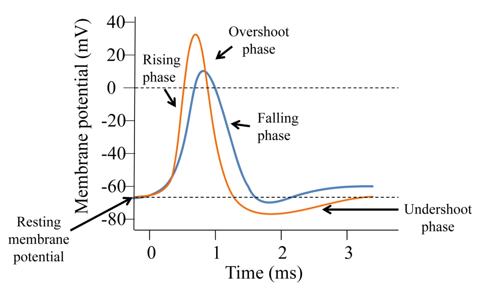
Fig.4.1 - Action Potential[link]
The Generation of an Action
- Methods of recording action potential
- intracellular -- smaller size
- extracellular
- Process
- Simulate
- The membrane of the nerve fibers is stretched
- Na+ permeable channels open
The Action Potential, In Theory
- There are three types of protein molecules: sodium-potassium pumps, sodium pumps, potassium pumps
Driving Force is equal to the real membrane potential and the equilibrium potential, which can be written as Vm-Ek(Vm is the membrane potential, Ek is the potential inside)
- $I_K=g_K(V_m-E_K)$
- More generally, it is writen as $I_{ion} = g{ion}(V_m-E{ion})$
At rest: $g_K > g_{Na}$
Ins: $g_{Na} >> g_K$(risingg edge) -- inward sodium current
Outs: $g_K >> g_{Na}$(falling edge) -- outward potassium current
Simply,switching the dominant membrane permeability from K to Na, the potential could be reversed rapidly.
The Action Potential, In Reality
Difficult to measure the sodium and potassium conductance of the membrane – Breaking techniques: Voltage clamp
The Voltage-Gate Sodium Channel
The protein forms a pore in the membrane is highly selective to Na+, and the pore is opened and closed by changes in membrane voltage
- Sodium Channel Structure
- polypetide:多肽
- It is for distinct domains numbered Ⅰ- Ⅳ, each domain consists of six transmembrane alpha helices.
- The four domains clump together to form a pore between them
- pore loop are selectivity filter
- S4 is voltage sensor
- the domains may arrange themselves to form a pore between them
- measurement function : patch clamp 膜片钳
- If the membrane potential does not reach threshold;potential, the change of potential have little effect on it.
Voltage-gated Potassium Channels
Different from sodium channels, it did not open immediately. There is a delay. It is called delay rectifier 延迟整流器
Putting the Pieces Together
Threshold: Membrane potential at which enough voltage-gated sodium channels open so that the relative ionic permeability of the membrane favor sodiums over potassium.
- Rising Phase: Na+ rushed into the cell, causing the membrane to depolarize.
- Overshoot: membrane potential goes closely to $E_{Na}$.
- Falling Phase: sodium channels inactivate. Potassium channels open.
Undershoot: The open voltage-gated potassium channels add to the resting potassium membrane permeability.
- Absolute refractory period: period when the channel inactivate and the channel is about to open.
Relative refractory period: the membrane potential stay hyperpolarized until the voltage-gated potassium channels close.
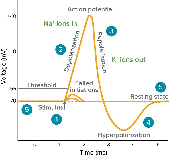
Fig.4.2 - Molecular basis of the action potential[link]
Action Potential Conduction
- soma to the axon terminal: orthodromic conduction -- without decrement
- backward propagation(either end of Axon couldd be depolarized)
- Action potential conduction velocities vary. But typically it is 10m/sec
- Influencing factors
The Farther the current goes down the axon, the farther ahead of the action potential the membrane will be depolarized, the faster the action potential will propagate; action potential conduction velocity increases with increasing axonal diameter.
Axonal size and the numbers of voltage-gated channels affect axonal excitability. smaller axons require greater depolarization to reach action potential threshold and are more sensitive to being blocked by local anesthetics.
- Myelin and Saltatory Conduction
- myelin 髓磷脂: wrapping the axon with insulation -- increase action potential conduction.
- There are breaks in the insulation where ions across the membrane to generate action potentials.
node of Ramvier(potential skip from node to node) -- this kind of potential is called saltatory conduction.
Action Potential, Axons and dendrites
- Dendrites and cell bodies do not generate sodium-dependent action potentials( very few channels).
- The axon hillock is ofter called spike-initiation zone. occurs near nerve endings.
- Aspinous stellate cells: typically responds to a steady firing rate.
- Spiny pyramidal cells: fire rapidly at the beginning and slow down.
Chapter 5 Synaptic Transmission
Synaptic transmission突触传递: information transfer at a synapse
Type of synapses
Electrical Synapse
- Allow direct transfer of ionic current from one cell to the next
it occurs at gap junctions (between cells in nearly every part of the body and interconnect many non-neural cells, some glandular cells 腺细胞 and glia)
At a gap junction, there are only 3 nm between the separated cells. The gap is spanned by clusters of special proteins called connexins 连接蛋白
- 20 subtypes of commexins -- half occurs in brain
- Six connexin subunits combine to form a channel called connexon
Two connexons(from each cell)meet and combine to form a gap junction channel -- the channel allows ions to pass directly from the cytoplasm of one cell to the cytoplasm of the other.
The pore of the gap junction is relatively large. -- big enough for all the major cellular ions and many small organic molecules.
- Gap junctions allow ionic current to pass equally well in both directions
Electrical Synapse is bidirectional. Because electrical current can pass through these channels, cells connected by gap junctions are said to be electrically coupled (fast -- enables an animal to beat a hasty retreat when faced with a dangerous situation).
Postsynaptic potential(PSP)突触后电位 : when two neurons causes a small amount of ionic current to flow across the gap junction channels into the other neuron. This current causes an electrically mediated postsynaptic potential.
- The precise role of electrical synapse vary.
Chemical Synapse
The pre- and post-synaptic membrane at chemical synapses are separated by a synaptic cleft裂缝. The cleft is filled with a matrix基质 of fibrous extracellular protein纤维细胞外蛋白.
Many axon terminals contain larger vesicles囊泡.(Secretory Granules分泌颗粒; Dense-core Vesicles致密核心囊泡)
Dense accumulation of protein adjacent to and within the membranes on either side of the synaptic cleft are collectively called membrane differentiations. On the pre-side, it looks like pyramid. The pyramids and the membrane associated with them are the actual sites of neurotransmitter release, called active zones.
Postsynaptic density: the protein thickly accumulated in and just under the postsynaptic membrane(contains the neurotransmitter receptors, which convert the intercellular chemical signal into an intercellular signal).
CNS Chemical Synapses
Asymmetrical synapse/Gray's type Ⅰ synapses: postsynaptic side is thicker than that on the presyunaptic side(excitatory兴奋的)
- Symmetrical synapse//Gray's type Ⅱ synapses: similar thickness(inhibitory抑制性)
Neuromuscular junction
- Chemical synapse also occur between the axons of motor neurons of the spinal cord and skeletal muscle
- Fast and reliable.
Principles of Chemical Synaptic transmission
Neurotransmitters
- amino acides
- small organic molecules
- store in and release from synaptic vesicles
- amines
- small organic molecules
- store in and release from synaptic vesicles
- peptides
- large molecules
- store and release from secretory granules
- Different neurons in the brain release different neurotransmitters
Neurotransmitter Synthesis and Storage
- Different neurotransmitters are synthesized in different way
The synthesizing enzymes for both amino acid and amine neurotransmitters are transported to the axon terminal, where they are locally and rapidly direct transmitter synthesis
Once synthesized, the amino acid and amine neurotransmitters must be taken up by the synaptic vesicles (transporters)
Neurotransmitter Release
- Release is triggered by the arrive of an action potential
- The depolarization of the terminal membrane causes voltage-gated calcium channels
Exocytosis: the vesicles release their contents. The membrane of the synaptic vesicle fuses to the presynaptic membrane at the active zone, allowing the contents of the vesicle to spill out into the synaptic cleft.
Neurotransmitter Receptors and Effectors
Membrane-spanning proteins consisting of four or five subunits that come together to form a pore between them.
- Same neurotransmitter can have different postsynaptic actions.
EPSP(excitatory postsynaptic potential): a transient postsynaptic membrane depolarization caused by excitatory. Synaptic activation os ACh-gated and glutamate-gated ion channels causes EPSPs.
Inhibitory postsynaptic potential(IPSP): an transient hyperpolarization of the postsynaptic membrane potential caused by the presynaptic release of neurotransmitter.
- G-Protein-Coupled Receptors
- All three types of transmitter all act on this.
- Reptor poteins embeded in the postsynaptic membrane.
The receptor proteins activate small proteins, called G-proteins(free to move along intracellular face of the postsynaptic membrane)
The activated G-protein activate "effector" proteins -- Second Messengers(it can activate additional enzymers in cytosol.)
Presynaptic receptors that are sensitive to the neurotransmitter released by the presynaptic terminal.
Autoreceptors are G-protein-coupled receptors that stimulate second messenger formation.
Neurotransmitter Recovery and Degradation
Once the neurotransmitter has interated, it must be cleared from the synaptic cleft to allow another roung of synaptic transmission.
- Simple diffusion (occurs by the action of specific neurotransmitter transport protein)
- Enzymatic detraction in the synaptic cleft(ACh)
Neuropharmacology
the study of drugs on nervous system
Principle of Synaptic Integration
The Integration of EPSPs
- Quantal Analysis of EPSPs
EPSP is some multiple of the response to the contents of a single vesicle(the total amount of transmitter released is some multiple of the contents of a single synaptic vesicle)
- Miniature Postsynaptic Potential: generated by the trasmitter contents of one vesicle
- EPSP Summation
- EPSP Summation: represents the simplest form of synaptic integration in the CNS
Spatial Summation: adding together of EPSPs generated simultaneously at many different synapses on a dendrite
Temporal Summation: adding together of EPSPs generated at the same synapse if they occur in rapid succeesion
The Contribution of Dentritic Properties to Synaptic Integration
The effectiveness of an excitatory synapse in triggering an action potential depends on how far the synapse is from the spike-initiation zone and ont the properties of the dendritic membrane
- Dendritic Cable Properties
- length constant: $V_{\lambda}=0.37(V_o)$
The length constant is an index of how far deporization can psread down a dendrite or axon
- Decrease in the end
Internal resistance: the resistance to current flowing longitudinally down the dendrite (depends on the diameter of the dendrite and the elctrical properties of the cytoplasm -- relatively a constant)
Membrane resistance: the resistance to current flowing across the membrane (depends on the number of open ion channels, which changes from moment to moment depending on what other synapses are active)
Excitable dendrites: The dendrites's membrane is electrically passive, lacks voltage-gated channels.
- Inhibition
- IPSPs and shunting Inhibition
The only important differences are that they different neurotransmitters and allow different ions to pass through their channels
Shunting inhibition: synapse acts as ame electrical shunt, preventing the current from flowing through the soma to he axon hillock (reduce the size of EPSPs, making the postsynaptic neuron less likely to fire actioin potentials)
- The Geometry of excitatory and Inhibitory Synapses
Inhibitory synapses use GABA or glycine as a neurotransmitter have a morphology charateristic of Gray's type Ⅱ -- contrasts with excitatory synapses that sue glutamate which have a Gray's type Ⅰ morphology.
Inhibitory synapses on many neurons are foudn clustered on the soma and near the axon hillock (powerful position to influence the activity of the postsynaptic neuron)
- IPSPs and shunting Inhibition
Modulation
many synapses with G-protein-coupled neurotransmitter receptors that are not directly associated with an ion channel (insteead modifies the effectiveness of EPSPs generated by other synapses with transmitter-gated channels)
- The norepinephrine beta receptor去甲肾上腺素β受体
Neurotransmitter norepinephrine(NE) to the βreceptor triggers a cascade of biochemical events within the cell -- activate a G-protein/ an effector protein, the intracellular细胞内的 enzyme adenylyl cyclase腺苷酸环化酶
Adenylyl cyclase 腺苷酸环化酶 catalyzes the chemical reaction that converts ATP. Its product of oxidative metabolism in the mitochondria 线粒体 into a compund called cyclic adenosine monophosphate(cAMP) 环磷酸腺苷
The effect of cAMP is to stimulate another enzyme know as a protein kinase蛋白激酶, which catalyze a chemical reaction called phosphorylation 磷酸化
Decreasing the K+ conductance increases the dendritic membrane resistance and therefor increases the length constant -- distant or week excitatory synapses will become more effective in depolarization the spike -initiation zone beyond threshold; cell become more excitable
Chapter 6 Neurotransmitter Systems
Studying Neurotransmitter Systems
Identifying the neurotransmitter:
- the molecule must be synthesized and stored in the presynaptic neuron
- The molecule must be released by the pesynaptic axon terminal upon stimulation
The molecule must produce a response in the postsynaptic cell that mimics the response produced by the release of neurotransmitter from the presynaptic neuron.
Localization of transmitters and transmitter-synthesizing enzymes
Immunocytochemistry 免疫细胞化学: when the same techniques applied to the brain, it is often referred as immunohistochemistry
- Neurotransmitter candidate has been chemically purified
- Injected to the skin and blood stream
- Stimulate an immune response
generation of a larger protein: antibodies (the best antibodies for immunocytochemistry bind very tightly to the transmitter of interest and bind very little or not at all to other chemicals in the brain)
- These antibody could be recovered and tagged with colorful maker.</p>
By using different antibodies, each labeled with a different marker color. It is possible to distinguish several types of cells in the same region of the brain.
- It can be used to localize any molecule for which a specific antibody can be generated
- In Situ Hybridization 原位杂交
- Process
- label appropriate probe --complementary strand
- apply it to a section of brain tissue
- wait
- wash away all the extra probes that have not stuck
- search for the label
- Chemically tagged
- radioactive & digital electronic image devices(autoradiography)
- label the probes with brightly colorful fluorescent molecules (FISH)
- It can be used to view the location of specific molecules
- Process
Studying Transmitter Release
- Impossible to stimulate a single population of synapses containing only a single neurotransmitter
- use brain slice that are kept in vitro 体外保存
- optogenetics -- activate one type of synaptic at one time
- We still not sure if molecules collected in the fluids were released from the axon terminals
Studying Synaptic Mimicry 突触模拟
- microiontophoresis 微离子电渗疗法: to assess the postsynaptic actions of a transmitter candidate
- Process
- a glass pipette 移液器 with a very fine tip, is filled with the ionized solution
- the tip is carefully placed next to the postsynaptic membrane of the neuron
- the transmitter candidate is ejected in a very small amounts by passing electrical current through the pipette
a microelectrode in the postsynaptic neuron can be used to measure the effects of the transmitter candidate on the membrane potential
If iontophoretic or pressure application 分子的离子电渗或压力应用 of the molecule causes electrophysiological changes 电生理变化 that mimic the effects of transmitter released at the synapse, and if the other criteria of localization, synthesis, and release are met, then the molecule and the transmitter are usually considered to be the same chemical.
Receptors
rule: no two neurotransmitter can bind to the same receptor. but one neurotransmitter can bind to many receptor
- neuropharmacological analysis of synaptic transmission
- two receptor could be distinguish by different drugs (different response) [nicotine ACh receptors & muscarinic ACh receptors]
- use selective antagonists 选择性拮抗剂 [curane inhibits the action of ACh at nicotinic receptors]
Different drugs are also used to distinguish several types of glutamate receptors 谷氨酸受体 [AMPA receptors, NMDA receptors, kainate receptors -- is activated selectively by a different agonist 激动剂]
- Ligand-binding methods
- many drugs interact selectively with receptors [Opiates, endorphins]
- Ligand 配体 for that receptors: And chemical compound that binds to a specific site on a receptor
- A ligand can be a agonist, an antagonistm or the chemical neurotransmitter itself
- molecular analysis of receptor proteins
two groups of neurotransmitter receptor proteins: transmitter-gated ion channels and G-protein-coupled(metabotropic) receptors
[GABA_A receptor have five subunits and give five major classes of subunit proteins,six different polypeptides]
Neurotransmitter Chemistry
- Dale's Principle: A neuron has only one neurotransmitter
- Co-transmitters: when two or more transmitters are released from one nerve terminal
Cholinergic Neuron 胆碱能神经元
Acetylcholine(ACh)乙酰胆碱 is the neurotransmitter at the neuromuscular junction and is therefore synthesized by all the motor neurons in the spinal cord and brain stem.
Its synthesis requires a specific enzyme, choline acetyltransferase(ChAT) 胆碱乙酰转移酶. ChAT is manufactured in the soma and transported to the axon terminal. Only cholinergic neurons contain ChAT
The transport of choline into the neuron is said to be the rate-limiting step in ACh sythesis
- AChE: Acetylcholinesterase 乙酰胆碱酯酶
Pumping Ions and Transmitters
The hard job of transporters is to pump transmitter molecules across membrane so effectively(concentrate in very small places)
Membranes of synaptic vesicles have pumps that us ATP to fuel the transport of $H^+$ into vesicles.
Transportaers use transmembrane gradient of $Na^+$ or $H^+$ as an energy source for moving transmitter molecules up steep concentration gradients.
The transporters themselves are large proteins that span membranes.Several transporters serve as a transmitters.
Plasma membrane transporters use a contransport mechanism carrying two $Na^+$ ions along with one transmitter molecule
Vesicular membrane transporters use a countertransport mechanism that trades a transmitter molecule from tje cytosol for a $H^+$ that trades a transmitter molecule from the cytosol for a $H^+$ from inside the cesicle.
Many psychoactive drugs such as amphetamines and cocaine, potently blocck certain transporters.
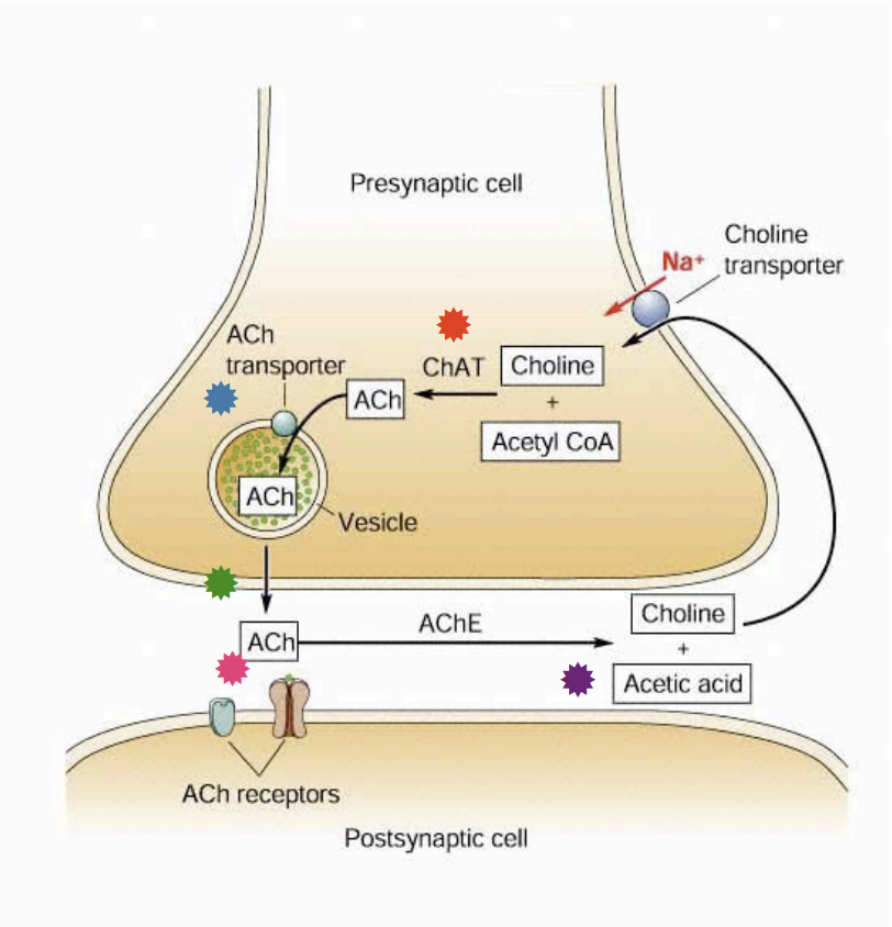
Fig 6.1 - The life cycle[link]
Catecholaminergic Neurons 儿茶酚胺能神经元
- neurotransmitters with catechol 儿茶酚 is called catecholamines 儿茶酚胺
The catecholamine neurotransmitters are Dopamine(DA) 多巴胺 , Norepinephrine(NE)去甲肾上腺素 , Epinephrine 肾上腺素/Adrenaline
- This process have no fast extracellular degradative enzyme analogous to AChE.
- The process is sensitive to cocaine and monoamine oxidase.
Serotonergic Neurons 血清素能神经元
- The amine neurotransmitter serotonin, also called 5-hydroxytryptamine(5-HT)
- The process is sensitive to MAO and antidepressant and antianxiety
Amino Acidergic Neurons 氨基酸能神经元
glutamate(Glu), glycine(Gly) and gamma-amino-butyric acid(GABA) 谷氨酸(Glu)、甘氨酸(Gly)和γ-氨基丁酸(GABA)
- Glutamate and glucine are synthesized from glucose and other precursors
Other Neurotransmitter Candidates and Intercellular Messengers
Adenosine Triphosphate(ATP) is also a neurotransmitter. It is concentrated in all synaptic vesicles in the CNS and PNS, and it is released into the cleft by presynaptic spiles in a $Ca^{2+}$ dependent manner.
- Endocannabinoids 内源性大麻素
They are not packaged in vesicles like most other neurotransmitters, instead, they are manufactured rapidly and on demand.
They are small and membrane permeable; once synthesized,, they can diffuse rapidly across the membrane of their cell of origin to contact neighboring cells.
hey bind selectively to the CB1 types of cannabinoid receptor, which is mainly located on certain presynaptic terminals.
When a postsynaptic neuron is very active, it releases enndocannabinoids, which suppress wither the inhibitory or excitatory drive onto the neurons.
- Nitric oxide(NO)
Transmitter-Gated Channels
A single channel can be a sensitive detector of chemicals and voltage, it can regulate the flow of surprisingly large currents with great precision, it can sift and select between very similar ions and can be regulateed by other receptor systems.(11nm long)
The Basic Structure of Transmitter-Gated Channels
Most contain the four hydrophobic segments that span the membrane in subunits of the nicotinic ACh recptor, the GABA_A receptor, and the glycine recepter. These three receptors are all pentameric complexes of subunits.
The glutamate-gated channels are slightly different. Glutamate receptors are tetramers, having four subunits that comprise a functional channel.
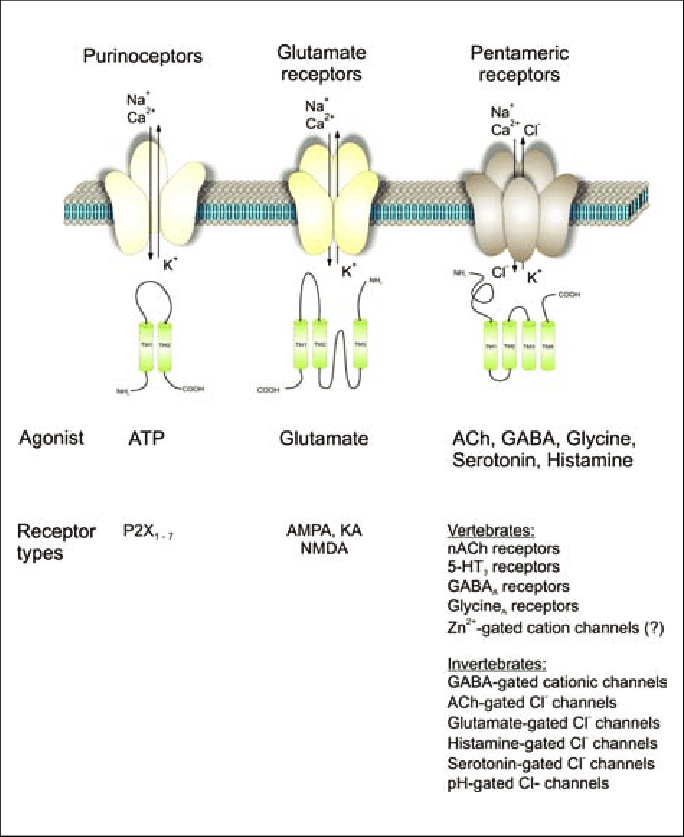
Fig 6.2 - Similarities in the structure of subunit for different transmitter-gated ion channels[link]
Amino Acid-Gated Channels
- Amino acid-gated channels mediate most of the fast synaptic transmission in the CNS.
- Several Propoerties of there channels distinguish them from one another:
The pharmacology of their binding sites decribes which transmitters affect them and how drug interact with them.
The kinetics of the transmitter binding process and channel gating determine the duration of their effect.
Thhe selectivity of the ion channels determines whether they produce excitation or inhibition and whether $Ca^{2+}$ enters the cell in significant amounts.
- The conductance of open channels helps determine the magnitude of their effects.
- Glutamated-Gated Channels
| glutamate receptor subtypes | AMPA | NMDA | Kainate |
|---|---|---|---|
| Ion permeable | $Na^+,K^+$ | $Ca^{2+}$ | $Na^+,K^+$(slower) |
| Potential | active at normal negative membrane | voltage dependent(when AMPA at the same and neighboring synapse) | ? |
| Function | mediate excitoatory transmission in much the same way as nicotinic receptors mediate synaptic excitation at neuromuscular juncion | Cause widespread and lasting changes in the postsynaptic neuron. | more than just an ionotropic |
- GABA-Gated and Glycine-Gated Channels
- GABA mediates most synaptic inhiition in CNS, and glycine mediates most of the rest.
Synaptic inhibition must be tightly regulated in the brain. Too much causes a loss of consciousness and coma; too little leads to a seizure.
G-Protein-Coupled Receptors And Effectors
- There are multiple subtypes of G-protein-coupled receptors in every known neurotransmitter system.
- Transmission at these receptors involves three steps: (1) binding of the neurotransmitter to the receptor protein, (2) activation of G-proteins, and (3) activation os effector systems.
The Basic Structure of G-Protein-Coupled Receptors
- Most receptors consists of a single polypeptide containing seven membrane-spanning alpha helices.
- Two of the extracellular loops of the polypeptide form the transmitter binding sites.
Structural variations in thie region determine which neurotransmitters, agonists, and antagonists bind to the receptor, sonsequently. which effector systems are activated in response to transmitter binding.
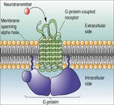
Fig 6.3 - Similarities in the structure of subunit for different transmitter-gated ion channels[link]
The Uniquitous G-Proteins
There are many more transmitter receptors than G-protein , so many types of G-proteins can be activated by many receptors.
Each G-protein has three subunits, termed $\alpha \ \ beta \ \ gamma$. In the resting state, a guanosine diphosphate(GDP) molecule is bound to the $G_{\alpha}$ sub-unit, and the whole complex floats around on the inner surface of the membrane.
If this GDP-bound G-protein bumps into the proper type of receptor and if thaat receptor has a transmitter molecule bound to it, then the g-protein releases its GDP and exchanges it for a GTP that it picks up from the cytosol.
The activated GTP-bound G-protein splits into two parts: the $G_{\alpha}$ subunit puls GTP and the $G_{\beta \gamma}$ complex. Both can then move on the influence various effector proteins.
The $G_{\alpha}$ subunit is itself an enzyme that eventually breaks down GTP into GDP. Therefore, $G_{\alpha}$ eventually terminates its own activity by converting the bound GTP to GDP.
The $G_{\alpha}$ and $G_{\beta \gamma}$ subunits come back together, allowing the cycle to begin again.
G-Protein-Coupled Effector Systems
The activated G-proteins exert their effects by binding to either of two types of effector proteins: G-protein-gated ion channels and G-protein-activated enzymes.
- A variety of neurotransmitters use the short-cut pathway, from receptor to G-protein to ion channel. One example is the muscarinic receptors in the heart.
- Shortcut pathways are the fastest of the G-protein-coupled systems, having responses beginning within 300-100 msec of neurotransmitter bind.
- The shortcut pathway is also very localized compared with other effector systems. Beacuase all the action in the shortcut pathway occurs within the membrane, it is sometimes called the membrane-delimited pathway
- G-proteins can also exert their effects by directly activating certain enzymes(second messenger cascade). Activation of these enzymes can trigger an elaborate series of biochemical reactions, a cascade that often ends in the activation of other "downstream" enzymes that alter neuronal funtion.
- synaptic transmission using transmitter-gated channels is simple and fast. Transmission involving G-protein-coupled receptors is complex and slow.
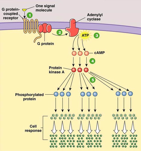
Fig 6.4 - Signal Amplification by G-protein-coupled second messenger cascades.[link]
Givergenve and Convergence In Neurotransmitter Systems
- divergence: The abilit of one transmitter to activate more than one type of postsynaptic response.
- Divergence is the rule among neurotransmitter systes,.Because of the multiple receptor subtypes, one transmitter can affect different neurons in very different ways.
- Divergence may occur at any stage in the cascade of transmitter effects.
- Neurotransmitters can also exhibit convergence of effects. Multiple transmitters, each activating their own receptor type, can converge to influence the same effector system.
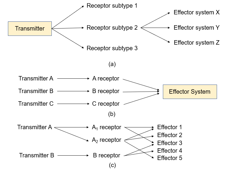
Fig 6.5 - Divergence and convergence in neurotransmitter signaling systems.
Conclusion
- Neurotransmitters are the essential links between neurons, and between neurons and other effector cells.
- Transmitters is one link in a chain of events, inciting chemical changes both fast and slow, divergent and convergent.
Chapter 7 The Structure of the Nervous System
Introduction
- Nervous system structure is the basis of understanding brain function.
- The human brain appears complicated because it is distorted as a result of the selective growth of some parts within the confines of the skull.
Gross Organization of The Mammalian Nervous System
The nervous system of all mammals has two divisions: the central nervous system(CNS) and the peripheral nervous system(PNS).
Anatomical References
- Anatomical References: reference within the brain.
- Anterior/Rostral : pointing toward the rat's nose( Latin: beak)
- Posterior/Caudal: pointing toward the rat's tail( Latin: tail)
- Dorsal: pointing up ( Latin: back)
- Ventral: pointing down ( Latin: bely)
- Bilateral symmetry: the right side of the brain and spinal cord is the mirror image of the left side.
- Midline: the invisible line running down the middle of the nervous system
- Medial: the structure close to midline
- Lateral: the structure far away midline
- Ipsilateral: two structure that are on the same side
- Contralateral: the structure are on opposite sides of the midline
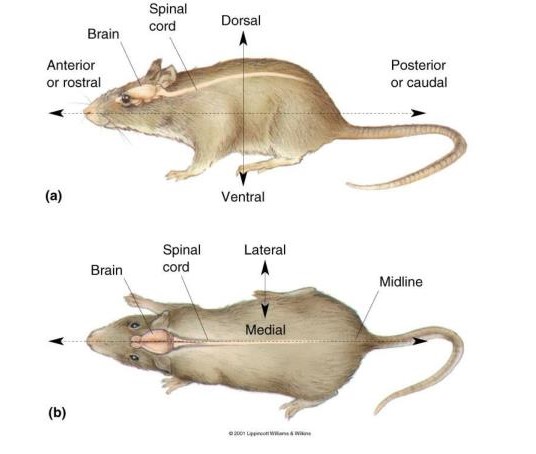
Fig 7.1 - Basic Anatomical references in the nervous system of a rat.(a)side view (b) top view.[link]
- In the language of anatomists, a lice is called a section; to slice is to section
- Midsagittal plane: splitting the brain into equal right and left halves
- Sagittal plane: Sections parallel to the midsagittal
- Horizontal plane: parallel to the ground
- Coronal plane: perpendicular to the ground and to the sagittal plane
- The anatomical planes are perpendicular to each other.
The Central Nervous System
- The central nervous system(CNS) consists of the parts of the nervous system that are encases in bond: the brain and the spinal cord.
- Cerebrum: the rostral-most and largest part of the brain. The middle plane split down the middle into two cerebral hemispheres, separated by the deep sagittal fissure. In general, the righ cerebral hemisphere receives sensations from, and controls movements of, the left side of the body. Similarly, the left cerebral hemisphere is concerned with sensations and movements on the right side of the body.
- Cerebellum: lying behind the cerebrum. The cerebellum is primarily a movement control center that has extensive connections with the cerebrum and the spinal cord. In contrast tp the cerebral hemispheres, the left body and the right side of the cerebellum is concerned with movements of the right side.
- Brain stem: the brain stem forms the stalk from which the cerebral hemispheres and the cerebellum sprout.The stem is a complex nexus of fivers and cells that relay information from the cerebrum to the spinal cord and cerebellum, and vice versa.
- Spinal Cord: the spinal cord is encased in the bony vertebral column and is attached to the brain stem. The spinal cord commmunicates with the body via the spinal nerves(part of the peripehral nervous system) Each spinal nerve attaches to the spinal cord by means of two branches, the dorsal root and the ventral root.
The Peripheral Nervous System
- All the parts of the nervous system otherthan the brain and spinal cord comprise the peripheral nervous system(PNS). The PNS has two parts: the somatic PNS and the visceral PNS.
- The Somatic PNS: all the spinal nerves that innervate the skin, the joints, and the muscles that are under voluntary contraol are part of the somatic PNS. The somatic sensory axons(innervate and collects information from the skin, muscles, and joints) enter the sinal cord via the dorsal roots. The cell bodies of these neurons lie outside the spinal cord in clusters called dorsal root ganglia.
- The Visceral PNS(Autonomic nervous system ANS): consists of the neurons that innervate the internal organs, blood vessels and glands. When one speaks of an emotional reaction that is beyond voluntary control(butterflies in the stomach), it usually is mediated by the visceral PNS.
- Afferent and Efferent Axons : indicate whether the axons are transporting information toward or away from a particular point.
The Cranial Nerves
- There are 12 pairs of cranial nerves that arise from the brain stem and innervate the head.
- Some of the cranial nerves are part of the CNS/somatic PNS/visceral PNS.
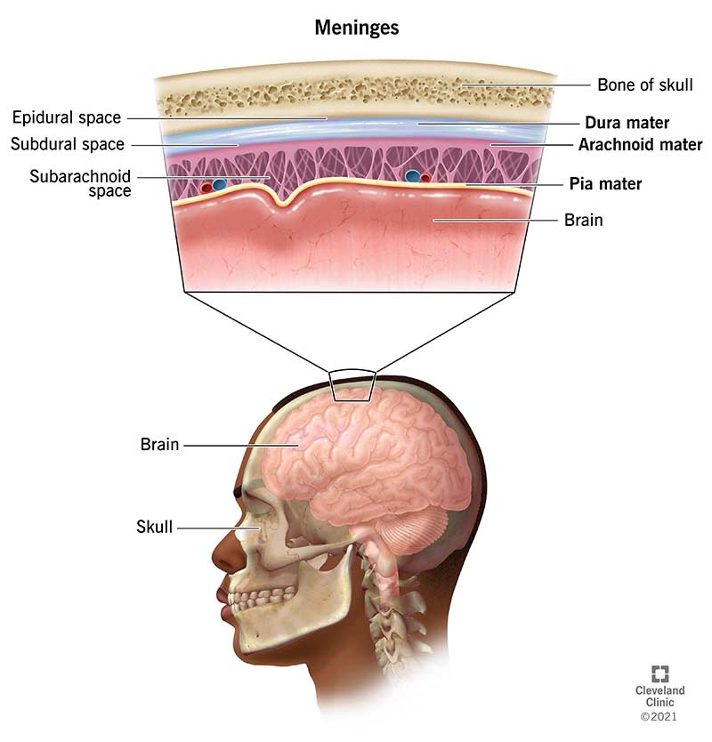
The Meninges
- The CNS is protected by thress membrane collectively called the meninges. The three membranes are the dura mater, the arachnoid membrane, and the pia mater.
- Dura mater: the outermost covering, forming a tough, inelastic bag that surrounds the brain and spinal cord.
- Arachnoid membrane: lie under the dura mater, with an appearance and a consistency resembling a spider web.
- Subdural hematoma: if the bood vessels passing through the dura are ruptured, blood can collect her and form what is called subdural hematoma. (normally no space between)
- Pia mater: a thin membrane that adheres closely to the surface of the brain. Many blood vessels ultimately dive into the substrate of the underlying brain along the pia.
- Cerebrospinal fluid(CSF): the pia is separated from the arachnoid by a fluid-filled space. </ul>
![]()
Fig 7.2 - The menings.[link]
#### The Ventricular System- </ul> ### Understanding CNS Structure Through Development ### A Guide To The Cerebral Cortex ### Conclusion ---
- </ul> test
- </ul> ### Understanding CNS Structure Through Development ### A Guide To The Cerebral Cortex ### Conclusion ---